
Detection method of sheep liver with B-ultrasound
Firstly, the sheep are held in a standing or supine position, and shearing is performed on the right side of the sheep at the level of the liver area 8-10 intercostal shoulder joints detected by B-ultrasound, so that the skin of the sheep’s liver body surface is detected by B-ultrasound revealed. First use the rechargeable electric shears to push away the thick and long hairs, then reduce the spacing density of the combs to cut off the remaining stubble. If necessary, apply an appropriate amount of coupling agent, then shave the hairs with a scalpel, and finally wipe the hair after applying the mixture. check.
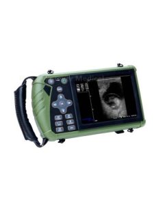
The B-ultrasound probe for sheep is kept in full and close contact with the skin. Care should be taken to avoid human-induced activities of the B-ultrasound probe for sheep and harassment of sheep. After scanning typical B-ultrasound images for sheep, freeze and save them, and record the scanning and storage time of the tomographic images. . After the B-ultrasound image of the sheep is frozen in time, the relevant indexes are measured. The audiogram stored in the B-ultrasound mainframe for sheep can be transmitted to the computer via infrared ray for storage by software.
Calcified hepatic echinococcosis occurs on the liver of sheep. The echoes of the sheep liver parenchyma are uneven. The echoes of sheep in the liver area are light spots or spots with B-ultrasound scanning. The number varies with the course of the disease. , which is in great contrast to the echo of normal liver tissue, showing partial or complete calcification
In the B-ultrasound operation of sheep, we will find that it is relatively easy to shear wool sheep with rechargeable portable shearing shears. Portable B-ultrasound for sheep is easy to operate, high in efficiency, easy to detect a clearer image, when scanning, apply force evenly and perform horizontal and vertical sliding or fixed-point rotation scanning on the liver area. In the process of B-ultrasound detection for sheep, it was found that there were ribs blocking the detection area, especially when the sheep were thinner, the ribs were obviously exposed, thus affecting the complete scan of the liver area, and the B-ultrasound we used for sheep showed that Two-dimensional tomographic images, thus resulting in a small number of sections that are difficult to display, may affect the results of diagnosis.