
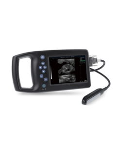
Knowledge of veterinary B-ultrasound probe
A veterinary ultrasound probe is a device that converts one form of energy into another. In ultrasound, sound waves are generated by electrical charges, causing a piezoelectric disk inside the probe to vibrate. Piezo is derived from the Greek word meaning “to press”. Piezoelectric materials are piezoelectric materials, such as quartz, that change shape and size when exposed to an electric field. Piezoelectric crystals are usually cylindrical, 1 to 2 cm wide and 1 mm thick. The current causes the crystal to expand and contract depending on the polarity of the current, and this vibration produces sound waves. The crystals also act as receivers, converting reflected sound waves into electricity, which in turn is converted into modified radio waves that produce images on the screen. Other piezoelectric substances include barium titanate, lithium sulfate, and lead zirconate. Barium titanate is most commonly used.
Sensors of various frequencies produce different penetrations. The frequency range is 1-25 MHz; frequencies of 3.5, 5.0, and 7.5 MHz are most common. High frequency probes produce short high frequency wavelengths (i.e. 7.5 MHz) and scan shorter distances with sharper resolution, low frequency probes produce longer wavelengths with greater beam penetration. Although these probes can scan at greater depths, the resolution is reduced.
Ultrasound is generated by the probe and directed towards the area of interest by holding the probe over the area. Some of the sound waves will be absorbed by the tissue and some will be reflected. The reflected part creates an “echo”, which is picked up by the probe. A homogeneous medium produces many echoes, which are echogenic regions, or no echoes, which are anechoic or anechoic regions. Tissues with poor echo, such as edematous tissue, have scattered echoes. Solid tumors are echogenic tissues producing many echoes at moderate sensitivities. Liquid is an anechoic medium and appears black on the screen, while bone is echogenic and appears white. Tissue away from the cystic (fluid-filled) area becomes hyperechoic because a large number of sound waves travel through the fluid.
Brightness-mode or B-mode systems use grayscale imaging to display echoes as points and display two-dimensional cross-sections of tissue. The second type of recording system, M mode or Sport mode, produces dots on the screen that are perceived as lines. Lines represent the distance from the reflex organ to the probe. The lines are usually recorded on strips of paper that move at a given speed. This trace is identical to the image on the screen and demonstrates motion, such as the contraction of the heart. Moving lines show the different positions of the valves, and the changing size of the heart chambers as the heart beats.
Current veterinary ultrasound systems use sector scanners or linear array probes. These “live” scanners watch movement by projecting continuous two-dimensional images. Sector scanners use one crystal of a given frequency mounted on a shaft that rotates once. Only part of the circle is open, sending and receiving signals in an arc. Therefore, the image seen is a portion of a fan-shaped cross-section of the tissue. Successive rotations and pulses allow for repeated imaging, displaying the area on the screen and allowing movement to be observed. Linear array probes use multiple crystals mounted in a straight line along the length of the probe. The crystals are individually pulsed sequentially to produce continuous scans to observe motion.