-
Breakthrough in China’s Medical Industry: Latest Advancements in Ultrasound Technology
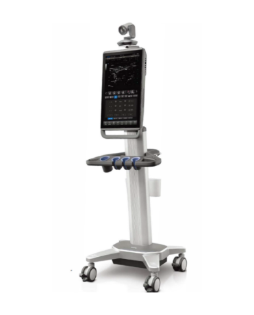
The medical industry in China is witnessing a remarkable breakthrough with the introduction of cutting-edge ultrasound technology. The latest advancements in this field are set to transform the landscape of medical imaging, enabling more accurate diagnoses, improved patient outcomes, and enhanced healthcare delivery. This news release provides an overview of the groundbreaking developments in ultrasound technology within the Chinese healthcare sector. High-resolution Imaging: Chinese researchers and manufacturers have made significant strides in developing ultrasound systems capable of producing high-resolution images. These systems utilize advanced transducer technologies and innovative signal processing algorithms to capture detailed anatomical structures with exceptional clarity. The improved image quality aids medical professionals in detecting subtle abnormalities and enables more precise diagnoses, leading to better treatment planning and patient management. Artificial Intelligence Integration: China is at the forefront of integrating artificial intelligence (AI) into ultrasound technology. By leveraging deep learning algorithms and machine learning techniques, AI-powered ultrasound systems can analyze vast amounts of imaging data in real-time. This integration enables automated image interpretation, the identification of specific pathologies, and the generation of quantitative measurements. The synergy between AI and ultrasound technology holds immense potential in enhancing diagnostic accuracy, reducing variability, and expediting the interpretation process. Point-of-Care Ultrasound...
-

The Functionality of Lead Aprons: A Comprehensive Explanation
Introduction: Lead aprons, commonly used in various fields such as medicine and radiology, are essential protective garments designed to shield individuals from potentially harmful radiation. With their unique properties and purpose-built design, lead aprons serve as a crucial barrier, minimizing the risks associated with exposure to ionizing radiation. This article aims to provide a comprehensive overview of the functionality of lead aprons, highlighting their key features and the benefits they offer. Radiation Shielding: The primary function of lead aprons is to provide effective radiation shielding. These aprons are constructed using a layer of lead or lead-equivalent material, which possesses excellent attenuating properties. When worn properly, lead aprons absorb and scatter ionizing radiation, preventing it from reaching the wearer’s body. This shielding capability is vital in environments where radiation exposure is a concern, such as X-ray rooms, nuclear medicine facilities, and interventional radiology suites. Protection of Vital Organs: Lead aprons are specifically designed to protect vital organs, such as the thyroid, breasts, and reproductive organs, which are particularly susceptible to radiation damage. The lead or lead-equivalent material used in the aprons effectively blocks the passage of ionizing radiation, safeguarding these sensitive areas from potential harm. By wearing lead aprons during medical...
-
Unveiling the Emerging Trends in China’s Healthcare Development in 2023
Introduction: As China’s healthcare sector continues to evolve rapidly, 2023 promises to be a year of pivotal transformation and innovation. With a focus on improving accessibility, quality of care, and leveraging cutting-edge technologies, the healthcare landscape in China is poised to witness several emerging trends that will shape the future of the industry. This press release highlights some of the key trends expected to dominate the healthcare development in China in 2023. Digital Health and Telemedicine Revolution: In 2023, China’s healthcare system will witness a surge in digital health solutions and telemedicine services. Leveraging advancements in artificial intelligence (AI), Internet of Things (IoT), and big data analytics, remote consultations, online diagnosis, and home-based healthcare monitoring will become more prevalent. These technologies will enhance accessibility to quality healthcare services, especially in rural areas, and provide efficient and convenient healthcare options for patients across the country. Expansion of Precision Medicine: Precision medicine, which tailors medical treatments to individual patients based on their genetic makeup and lifestyle factors, will continue to gain momentum in China. The country’s commitment to genomics research, coupled with the development of precision medicine initiatives, will enable more personalized and targeted therapies for various diseases. The integration of genetic...
-
An Introduction to Ultrasound-Guided Anesthesia
Ultrasound-guided anesthesia, also known as ultrasound-assisted regional anesthesia, is a technique that utilizes real-time ultrasound imaging to enhance the precision and safety of administering anesthesia. This approach has revolutionized the field of anesthesia by providing detailed visualization of anatomical structures and improving the accuracy of needle placement. In this article, we will explore the principles, benefits, and applications of ultrasound-guided anesthesia. Principles of Ultrasound-Guided Anesthesia: Ultrasound-guided anesthesia involves the use of a handheld ultrasound probe that emits high-frequency sound waves. These sound waves penetrate the body and bounce back to create images of the underlying structures, such as nerves, blood vessels, and muscles. By visualizing these structures in real-time, anesthesiologists can accurately guide the placement of needles and administer anesthesia with precision. Benefits of Ultrasound-Guided Anesthesia: Enhanced Safety: One of the primary advantages of ultrasound guidance is the improved safety it provides. By visualizing the target structures, the risk of accidental injury to adjacent tissues, nerves, and blood vessels is significantly reduced. Increased Accuracy: Ultrasound imaging allows anesthesiologists to precisely locate the target nerve or tissue for anesthesia administration. This accuracy reduces the likelihood of failed blocks or inadequate pain relief, leading to improved patient outcomes. Reduced Complications: Compared to...
-
China’s Healthcare Industry Sets Direction for Accelerated Development in Pharmaceutical and Medical Device Innovation
The healthcare industry in China is witnessing a remarkable shift towards accelerated innovation in pharmaceuticals and medical devices, as the nation sets its sights on becoming a global leader in healthcare advancements. With a clear vision and strategic initiatives, China’s medical sector is poised to revolutionize patient care, foster drug discovery, and boost technological breakthroughs in the years to come. China’s healthcare industry has made significant strides in recent years, propelled by the government’s commitment to improving healthcare accessibility, quality, and affordability for its vast population. This renewed focus has led to the formulation of a comprehensive development plan aimed at fostering innovation and propelling the nation’s medical sector to new heights. Under this strategic direction, pharmaceutical and medical device innovation has emerged as a key aspect of China’s healthcare agenda. Recognizing the need to address the growing demand for effective treatments and cutting-edge medical technologies, the Chinese government has implemented several measures to expedite the development and approval processes for new drugs and medical devices. One notable initiative is the streamlining of regulatory procedures, paving the way for faster approval timelines without compromising safety and efficacy standards. By implementing more efficient regulatory frameworks, China aims to attract global pharmaceutical...
-
The Function and Significance of Ultrasound Convex Array Probes
Introduction: Ultrasound imaging is a widely used medical imaging technique that utilizes sound waves to generate real-time images of internal body structures. Among the various components of an ultrasound system, the probe plays a crucial role in transmitting and receiving ultrasound waves. One type of probe commonly employed in medical settings is the convex array probe, renowned for its unique features and versatility. In this article, we will explore the functions and significance of ultrasound convex array probes. Functionality of Convex Array Probes: Convex array probes consist of multiple piezoelectric elements arranged in a curved or convex shape. These elements emit ultrasound waves and receive the echoes reflected back from the tissues. The curved design allows for a wider field of view, enabling the imaging of larger anatomical areas. The probe’s elements are electronically steered to create a focused beam, which enhances the image resolution at different depths. This capability makes convex array probes suitable for examining organs located deep within the body, such as the liver, heart, and abdomen. Wide Range of Applications: Convex array probes find applications in various medical specialties due to their versatility. They are particularly valuable in obstetrics and gynecology, where they allow for detailed...
If you have any question, please contact us