未分类

Home > 未分类

4D Doppler Ultrasound Machine Price

* Simplified Chinese, Traditional Chinese, English, French, Spanish, Japanese, Polish
* Position markers: About 125
* Printer Settings: video printer, report printer
* With automatic freezing function
* With foot switch function
* Settings Image storage format: JPG, BMP, DCM, TIF, PNG
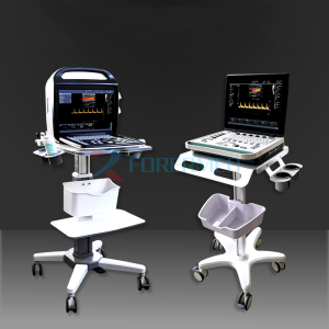
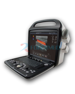
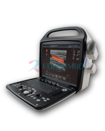
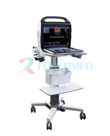
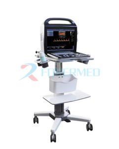
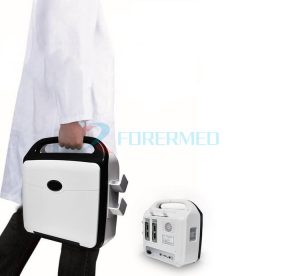
4D Color Doppler Ultrasound Scanner YJ-PR8

Features

1 Have black and white correlation Video output:
Support three color TV standards, NTSC, PAL, SECAM.
2 System Settings Interface language
* Simplified Chinese, Traditional Chinese, English, French, Spanish, Japanese, Polish
* Date format: The value can be YYYY-MM-DD, mm/dd/yyyy, mm.dd.yyyy, dd/mm/yyyy
* Time format: 24 hours
* Screen saver available
* Position markers: About 125
* Printer Settings: video printer, report printer
* With automatic freezing function
* With foot switch function
* Settings Image storage format: JPG, BMP, DCM, TIF, PNG
 
Preset application mode:
In B/CFM/PW mode, 26 groups of preset mode Settings can be performed respectively (including 8 groups of user-defined Settings)

Default text comment:
Each group of application modes can be set 10 text comment content (each text comment ** more than 15 bytes) Obstetrical table,
gestational age setting: set obstetrical measurement site, measurement method, view gestational age table and
fetal growth table, create a custom gestationalage table, fetal growth table with system upgrade, with maintenance tools.
 
3 Image Preprocessing
Total gain: adjustable TGC: 8 TGC regulating rod
Sound output: adjustable
Grayscale value: multilevel adjustable

4 Image Processing
Frame average: multilevel adjustable
Gray threshold: multilevel adjustable
Gamma correction: multilevel adjustable
Contrast adjustment range: adjustable
GIP: multilevel adjustable
Spatial composite switch: adjustable
Trace: multilevel adjustable
5 Measurement and calculation
* B-mode routine measurement: distance, circumference, area, volume, Angle, ratio,
stenosis rate, etc.
* M-mode routine measurement: heart rate, time, distance, slope, ratio, etc.
* Gynecological measurement: uterus, cervix, endometrium, ovary, follicle, etc.
* Obstetric measurement: gestational age estimation, due date, fetal weight estimation, amniotic fluid index
* Cardiac measurements: left ventricular measurements, etc
* Urology measurements: prostate volume, transitional area volume, bladder volume,residual urine volume, etc
* PW measurement: Doppler measurement, time measurement, velocity measurement,
heart rate measurement, RI measurement, PI measurement etc.
* Orthopaedic surgery measurements: cross section measurement, hip measurement, etc
* Vascular measurement: thickness of endovascular media, length stenosis ratio, area stenosis ratio

Technical specifications

7 Display mode: B mode, 4B mode, B/M mode, M mode (M extended), anatomy M super, CFM mode, PW mode, B/PW, D extended, dual
display, black and white/color dual real-time display mode

8 Image display: 

256 gray scale display

Image rotation: left/right, up/down, 90 degree rotation
Depth range: Each probe has a corresponding depth range

Transmission focusing: multiple focusing positions (number of focusing positions depends on probe type and depth)
Receiving focus: continuous dynamic focus, dynamic aperture
Dynamic range: adjustable
Real-time scaling: adjustable magnification
M mode speed: adjustable
Change perspective: adjustable
The TGC curve is displayed

System Configuration

Machine, display

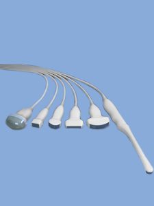
Convex array probe:

C3501, center frequency 3.5MHz, suitable for abdominal, gynecology, obstetrics, urology examination.

Linear array probe:

L7501, center frequency 7.5MHz, suitable for small organs, breast examination.

Transvaginal array probe:

E6501, center frequency 6.5 MHz, suitable for transvaginal and transrectal examination.

Phased array probe:

P2501, center frequency 2.5MHz, suitable for cardiac examination.

Patient information management software, tissue harmonic imaging software

Disk storage capacity:

2500 GB

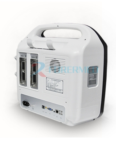
USB standard interface:

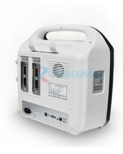
Supports USB2.0, 2 interfaces

Supports both black and white video printers and color video printers

DICOM3.0 transmission:

After the ultrasonic device is connected to PACS, the required image can be transmitted and obtained through the DICOM protocol.

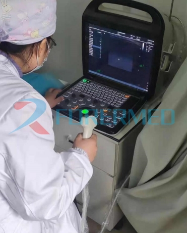
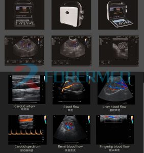
Clinical Image

Related Products

Send us a message

If you have any question, please contact us