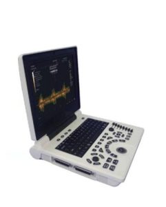
Introduction to Imaging—Basic Knowledge of Ultrasound
(1) Basic principles of ultrasound imaging
The vibration frequency of the sound source commonly used in ultrasonic diagnosis is 2.5~5.0MHz. When the ultrasonic wave propagates in the human body and passes through different organs, tissues, and lesions, the acoustic impedance of the medium on both sides of each interface is different, and different degrees of reflection, scattering, and ultrasonic attenuation occur. The information is obtained by the receiver, and after information processing, it is displayed as a waveform or image on the screen. According to the type and display mode of ultrasound imaging, it is divided into A-type ultrasound, B-type ultrasound (two-dimensional ultrasound), M-type ultrasound, and D-type ultrasound.

(2) Main features of 2D sonogram
①The imaging record is a two-dimensional cross-sectional view of the inspection site, and the inspection site and scanning direction are often marked.
②The image is composed of black, white, and gray dots, which represent the strength of the tissue structure echo, and the whiter the color, the stronger the echo (distinguishing between high-density and low-density X-ray and CT).
③The image display range is limited, and larger organs and lesions cannot be displayed as a whole.
④Contrast-enhanced acoustic examination changes the echo of the tissue structure on the image.
(3) Normal appearance of two-dimensional ultrasound images
1. Liver
The size and outline are the same as the previous examination, and the echo signal of the liver parenchyma is lower than that of the diaphragm, which is roughly equal to that of the pancreas.
2. Gallbladder
On the cross-section and longitudinal section, it is round, quasi-circular, or oblong, with a long diameter of less than 9cm, an anteroposterior diameter of less than 3.5-4cm, and a wall thickness of 2-3mm. The edge of the gallbladder wall is smooth and hyperechoic. Echo, increased echo in the back of the gallbladder.
3. Kidney
Round, oval, and bean-shaped, the capsule is smooth and clear, hyperechoic, the renal parenchyma is uniform and weakly echogenic, the renal pyramids are hypoechoic, and the ureter is often not displayed due to intestinal gas interference.
(4) Manifestations of liver lesions
In liver cirrhosis, ultrasound can directly detect liver atrophy, whole liver volume reduction, deformation, uneven surface, diffuse thickening and enhancement of echoes, uneven and dense small point-like abnormal echoes, thinning, tortuous, and stiffness of hepatic veins, and indirect manifestations. Including splenomegaly, peritoneal effusion, portal vein trunk and main branch thickening.
(5) Manifestations of gallbladder lesions
1. size
Shrunken gallbladder with thickened gallbladder wall is seen in chronic cholecystitis, and enlarged gallbladder indicates cholecystitis and biliary system obstruction.
2. Calcification
Stones in the gallbladder and bile duct manifest as strong echoes, accompanied by acoustic shadows behind, and gallbladder stones can move with changes in body position.
3. Expansion
The inner diameter of the intrahepatic bile duct is greater than 2 mm, and the inner diameter of the common bile duct is greater than 6 mm, suggesting dilatation.
4. Filling defect
Stones and tumors can cause filling defects in the cyst cavity, and the edges of stone defects are smooth, and tumors are mostly irregular.
(6) Manifestations of kidney disease
Ultrasound is highly sensitive to kidney stones, and can detect hyperechoic calyces, renal pelvis, or ureter in the form of dots or masses, with sound shadows visible behind, and dilated hydrops in the upper ureter and renal pelvis when obstructed.

