
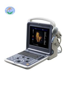
Popular science knowledge of ultrasonography
Ultrasound is to use the ultrasonic probe to repeatedly scan and receive the ultrasonic sound beam, and display the received different echo intensities on the corresponding position of the screen to outline the gray-scale section image. The received ultrasound is a high-frequency sound wave, which is safe and harmless to the human body. It is also very safe to be widely used in the examination of various organs of the body and the fetus. Among them, interventional ultrasound is also a minimally invasive treatment. Under the real-time dynamic monitoring and guidance of ultrasound, various operations such as puncture, biopsy, suction, intubation, drug therapy, microwave, and radiofrequency ablation are completed. The operation time is short, recovery is fast, It has the advantages of fewer complications and has been widely used in clinical practice.
Do Ultrasounds Have Radiation?
The “ultra” in ultrasonography refers to ultrasound, which is essentially the same as the sound you hear, but ultrasound is a special high-frequency sound wave that cannot be heard by the human ear, so there is no radioactivity, so you can check it with confidence. The advantages of ultrasonography, such as economy, convenience, and repeatability, have now become one of the first-choice inspection methods in clinical practice.
Why do some ultrasound examinations require fasting, and some examinations require holding back urine?
This is determined according to the physiological characteristics of the human body and the principles of ultrasound examination. After eating, the gallbladder will contract due to the emptying of bile, which will cause the gallbladder to be unclear, and the contents and gas in the gastrointestinal tract will affect the transmission and reflection of ultrasound in the human body, making the ultrasound image blurred; If the bladder is filled with fluid, it is like a deflated balloon. Ultrasound cannot detect diseases of the bladder. On the contrary, if the bladder is filled well, it can not only find the problems of the bladder itself, but also provide a sound-transparent channel for the uterine appendages or prostate around it. window for clearer and more reliable images.
What should I pay attention to before ultrasound examination?
- The digestive system (liver, gallbladder, spleen, pancreas), abdominal blood vessels, and retroperitoneal lymph nodes need to be fasted for 8-12 hours.
- Examinations of the urinary system (kidney, ureter, bladder, prostate), gynecology (uterus, appendages, early pregnancy, placenta previa, suspected cervical insufficiency, cesarean section scar conditions) and other examinations need to hold back urine to fill the bladder.
- Bladder should be emptied before transvaginal/rectal ultrasonography.
- Most ultrasonography examinations in children (especially echocardiography in children) need to be performed in a quiet state, and appropriate sedatives can be given by the pediatrician if necessary.
- Some ultrasound examinations (breast, scrotum, extremities, etc.) need to fully expose the examination site. It is recommended to wear loose, easy-to-wear and easy-to-take-off, plain clothing; for neck, limbs and other examinations, jewelry should be removed early for examination.
There are many inspections at the same time, what should be done first?
Ultrasonography should be scheduled before endoscopy (gastroenteroscopy), barium meal, and cholangiography. Ultrasound examinations for special items (such as interventional ultrasound, contrast-enhanced ultrasound, four-dimensional color Doppler ultrasound in second pregnancy, etc.) need to be booked in advance.
The scope of application of ultrasound
- Head and neck, brain, intraocular and orbital, maxillofacial, neck, thyroid, parathyroid, lymph nodes, etc.
- Chest, mammary gland, chest wall, chest cavity, lung and heart, etc. 3. Abdomen, abdominal cavity and retroperitoneum, liver, gallbladder, spleen, pancreas, kidney, gastrointestinal tract, uterine appendages, bladder, prostate, etc.
- Male reproductive system: scrotum, testis, epididymis, spermatic cord and seminal vesicles, etc.
- Abdominal great vessels and peripheral vessels, etc.
- Joints and soft tissues, etc.
Interventional Ultrasound Diagnosis and Therapy
- Puncture biopsy of tumors in the neck, thyroid, breast and other superficial parts and abdominal organ tumors, pathological diagnosis of benign and malignant.
- Histopathological diagnosis of diffuse liver and kidney lesions.
- Puncture, aspiration and drainage, drug flushing and sclerotherapy for fluid-containing lesions (liver, kidney, ovary, thyroid, breast cyst, abscess, and effusion, etc.).
- Catheter drainage for intrahepatic biliary tract and renal obstructive diseases, fetal amniocentesis, etc.
- Intratumoral (thyroid nodules, liver cancer, hepatic hemangioma, uterine fibroids, etc.) puncture drug therapy, ultrasonic positioning and guidance in microwave and radio frequency therapy, etc.