If you have any question, please contact us
If you have any question, please contact us
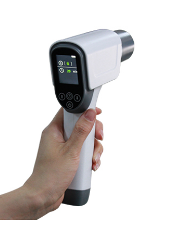
Abdomen, Obstetrics, Gynecology, Pediatrics,Small parts, Artery, Superficial organ, Orthopedics, Cardiology, Musculoskeletal, Vascular, etc
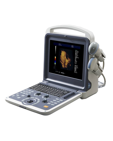
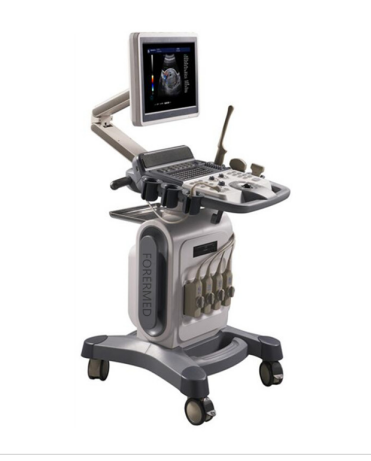
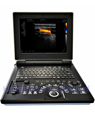
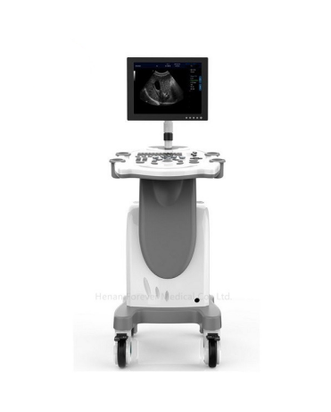
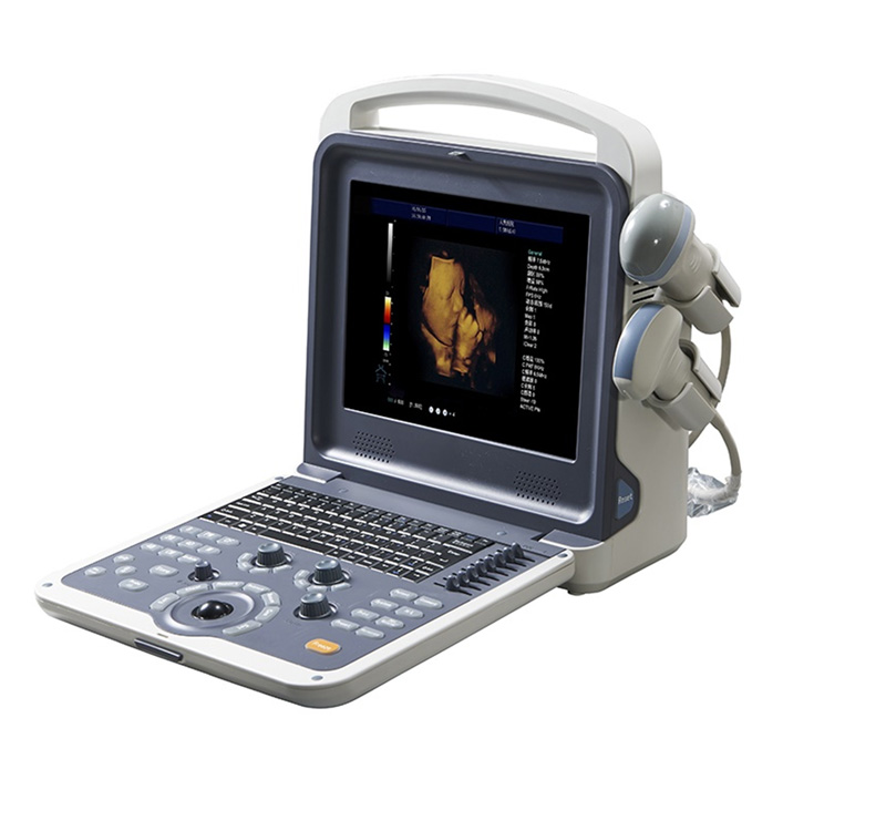
Full Digital Color Doppler Ultrasound Diagnostic System
|
Display mode |
- B, B|B,4B, B|M,M,B|D,PW,B|PW, CF - Duplex/Triplex mode - CW (option) - 4D mode (option)
|
|
Zoom
|
- Real time zooming - 10 Steps: ×1.0, ×2.0, ×3.0, ×4.0, ×5.0, ×6.0, ×7.0, ×8.0, ×9.0, ×10.0 - Selectable zooming position |
|
Focus |
- Continuous dynamic focus - 1~16 selectable transmit focus - Acoustic lens focus - 1, 2, 3, 4 focus
|
|
Memory Cine‐memory
|
- B‐mode - M‐mode - SSD (Solid State Disk) 64G |
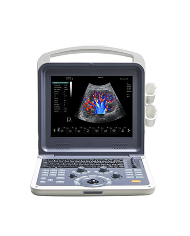
Appearance
- Smart, compact and clamshell design
- 12 inch LED monitor
- Backlit operation panel, 8TGC
- Floating keyboard
- Two active probe connectors
- Two probe holders
Probe
Transducer Types
- 3.5MHz convex probe (2.0/ 3.0/ 3.5/ 4.0/ 5.5MHz)
- 7.5MHz linear probe (6.0/ 6.5/ 7.5/ 10.0/ 12.0MHz)
- 3.5MHz micro convex probe (2.0/ 2.5/ 3.5/4.5/ 5.0MHz)
- 6.5MHz transvaginal probe (5.0/ 6.0/ 6.5/ 7.5/ 9.0MHz)
- 3.5MHz phased array probe (2.1/ 3.0/ 3.5/ 4.0/ 5.0MHz)
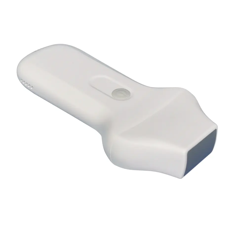
- 4D volume probe (2.0/ 3.0/ 3.5/ 4.0/ 5.5MHz)
Applications
- Abdomen, Obstetrics, Gynecology, Pediatrics,Small parts, Artery, Superficial
organ, Orthopedics, Cardiology, Musculoskeletal, Vascular, etc
Function
- Auto Image Optimization
- Tissue Harmonic imaging
- iClear (Speckle Noise Reduction)
- iBeam (Spatial Compound Image)
- iZoom
- PIHI (Pulse-Inverse Harmonics Imaging)
- SA (Synthetic Aperture ultrasonic Imaging)
- Panoramic Image (Option)
- Trapezoid Image (Option)
- Continuous Wave Doppler(Option)
Physical Features
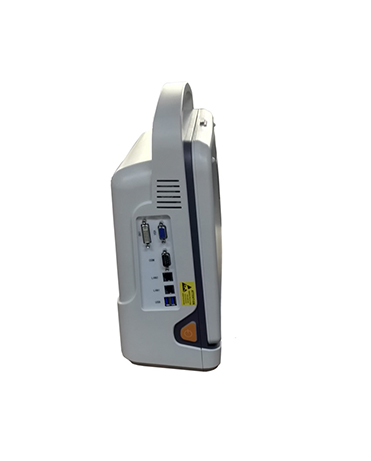
Connectivity
- Video out port
- DVI out port
- VGA out port
- 2 USB port
- DICOM 3.0
Dimension
- Gross dimension: 510 mm X 500 mm X 330mm
- Net dimension: 330mm X 150 mm X 380mm
Weight
- Gross weight : 12 kg
- Net weight : 7 kg
Power Requirements
- Voltage: AC 100V to 240V±10%
- Frequency: 50Hz±1Hz
- Rated Power: 250VA
Operation Conditions
- Ambient temperature: 0℃to +40℃
- Relative humidity: 38% to 85%
- Atmospheric Pressure: 700hPa to 1060hPa
Software & Accessories
Standard Accessories
- Power Cable
- Operation Manual
- Fuse
- System Recovery USB
- Built in Li‐ion battery
Optional Accessories
- B/W or color Video printer
- LaserJet or inkjet printer
- Trolley
- Aluminum case
Imaging Processing
|
B mode
|
- 8‐step TGC slide pots - Gain: 0~100% - Depth: 1.6~30cm - Frequency: 5 steps - Dynamic range adjustable: 0~150dB - Edge enhancement:0~7 - Persistence:0~7 - Chroma:0~6 - Grayscale:0~16 - Power: 0~100% - Noise reduction: 0‐6 - iclear: off, 1, 2, 3, 4 |
|
M mode |
- Gain: 0~100% - Sweep speed: 4 steps - Maps: 0~16 - Chroma:0~6 |
|
C mode |
- Gain: 0~100% - Pulse wave - Wall filter: 4 steps - Color Maps: 0~7 - Package size: 8~15 - Color persistence: 0~7 - Threshold: 0‐3 - Base line: 0‐6 - Line density: Low and high - Spatial filter: 0‐3
|
|
PW mode |
- Gain: 0~100% - Frequency: 5 steps - Pseudo color:0~6 - PRFd:1.0~6KHz - Basic line: 7 steps - Wall filter: 7 steps - Spectrum mode: Refresh and Synchronize |
Measurement & Calculation
Measurement
|
B mode (General)
|
- Distance - Trace Length - Ellipse (area) - Trace(area) - Angle - Volume |
|
PW mode
|
- HR (heart rate) - Distance - Velocity - Time |
Calculation
|
Abdomen
|
- Liver - Gallbladder - Pancreas - Spleen |
|
Urology
|
- Kidney - Ureter - Bladder - After the urine bladder - Prostate |
|
Gynecology
|
- Uterus - Cervix - Ovary - Follicle |
|
Early Obstetrics
|
- GS - BPD - CRL - NT … |
|
Later Obstetrics |
- BPD - HC - AC - OFD - FL - TAD ... |
|
Small parts |
- Thyroid - Testes |
|
Musculoskeletal |
- Hip |
|
Peripheral vascular
|
- Intima - Artery |
|
Cardiology
|
- Distance - Angle - Volume - RVWd - LVDd - RVDd - LVPWd - RVWs - LVDs - RVDs - LVPWs - RV/LV - AO … |
If you have any question, please contact us