-

Do you know the functional parameters of the multi-parameter monitor?
What are the functional parameters of the multi-parameter monitor? The monitor is a practical precision medical instrument in the hospital. It can simultaneously monitor the patient’s dynamic and practical precision medical instrument ECG, respiration, body temperature, blood pressure (non-invasive and invasive), blood oxygen saturation, pulse rate and other physiological parameters. 24-hour continuous monitoring of the patient’s physiological parameters, detection of changing trends, pointing out the critical situation, for doctors to deal with emergency treatment and the basis for treatment, to minimize complications, to achieve the purpose of alleviating and eliminating the disease. The standard six parameters of the monitor are ECG, respiration, non-invasive blood pressure, blood oxygen saturation, pulse, and body temperature. In addition, optional parameters include: invasive blood pressure, end-tidal carbon dioxide, respiratory mechanics, anesthetic gas, cardiac output (invasive and non-invasive), EEG bispectral index, etc. The clinical application scope of the monitor: during operation, after operation, trauma care, coronary heart disease, critically ill patients, neonates, premature infants, hyperbaric oxygen chambers, delivery rooms, etc.
-
What is the gel applied for ultrasound examination?
What is the gel applied for ultrasound examination? Health care workers who perform ultrasound examinations always apply a layer of clear gel before touching the probe with the examiner. You might think it’s a lubricant, but that’s just a secondary role. The official name of this gel is Ultrasonic Coupling Agent, and its main purpose, as its name suggests, is as a medium for “coupling”. Ultrasonography scans the human body with ultrasound waves (frequency greater than 20 kHz) that are inaudible to the human ear, and converts the echo signals into image information inside the body. B-ultrasound presents black and white images with different brightness (Brightness), while color Doppler ultrasound presents the blood flow of tissues in color on this basis. However, as a mechanical wave, the propagation of sound waves is affected by changes in the medium. Although the probe of the ultrasound equipment is next to the skin, the dry contact is inevitably separated by air. Due to the large difference in acoustic impedance between the air and the human body, two completely different media, sound waves will be reflected a lot at the interface between the two, and a lot of sound energy has been lost before...
-
What is Ultrasound Bone Densitometer?
Osteoporosis is not a serious disease in the eyes of many people, and it has not attracted everyone’s attention. This chronic disease may not cause death. Many people do not choose to test or seek medical treatment even if they know that they may have low bone density. The bone density test has already been planted in their hearts. It is a lie, and they don’t want to be fooled. Eating a little more good food and exercising can make up for it. Contemporary women, in the age group of 25 to 35, more than 50% of white-collar women have more severe bone loss than men, and the incidence is significantly higher than that of men. Women feel lower back pain, a considerable part of which is an early symptom of osteoporosis. Nowadays, many young women are prone to osteoporosis due to dieting to lose weight, sitting more and moving less, and unbalanced diet. Changes in bone mineral density during pregnancy and lactation are caused by the growth and development of fetuses and infants. In contemporary men, middle-aged men begin to lose bone mass due to smoking, alcoholism, and metabolic diseases such as obesity, diabetes, and hypertension. If you have...
-
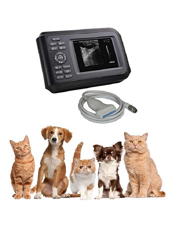
Role of vet ultrasound in animal pregnancy examination
Detailed function description of the veterinary ultrasound machine for animal pregnancy examination: 1. Check whether the animal is conceived successfully 2. Observe embryonic development: Judge the embryonic development by observing the changes of the fetus’s external structure and internal structure. 3. Monitoring the life and death of the fetus: Using ultrasound to detect the heartbeat of the fetus can predict the life and death of the fetus. Before the embryo dies, the heartbeat is significantly reduced. Fetal movement disappears, the fetal sac is filled with dark areas of liquid, the embryo cannot be seen, the echo in the uterus is disordered, and the fetal sac, placenta, and fetal structure cannot be distinguished, all indicating embryonic death. 4. Fetal heart rate and its pulsation: Fetal heart rate can be calculated by measuring fetal heart or fetal arterial pulsation (including umbilical cord artery-referred to as fetal blood sound). 5. Identify the sex of the fetus: Using ultrasound to detect the positional relationship between the reproductive structure of the fetus and the surrounding structures can accurately identify the sex of the fetus. 50 to 105 days after the cattle are bred, the accuracy of identifying the sex of the fetus is 96%. 6....
-
What The Advantages Of PCR Test
Digital PCR can directly measure the number of nucleic acid molecules in the initial sample, which is the absolute value quantification of nucleic acid concentration, and is an epoch-making nucleic acid detection technology. Compared with qRT-PCR, the core advantages of digital PCR are reflected in high detection accuracy, high sensitivity, simple overall operation, stable system, excellent repeatability, and can effectively avoid cross-contamination between samples, specifically: (1) The reported result is the copy number of the target gene, which provides a more accurate nucleic acid quantification basis for clinical diagnosis and treatment; (2) The detection gray area is greatly reduced. By intuitively reading the quantitative results, human errors are avoided, and the high reliability and repeatability of the detection results are guaranteed; (3) It can eliminate the interference of samples with complex components such as feces on the experimental results, and avoid false negative results caused by component inhibitors. In conclusion, digital PCR can effectively avoid the missed detection of patients with low viral load, and help to detect the disease as soon as possible and take isolation measures. Recently, the Frontier Center of the Chinese Academy of Metrology has successfully developed a SARS-CoV-2 nucleic acid detection reagent based on digital...
-
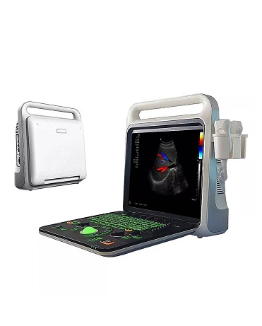
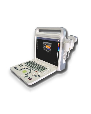
What is a color Doppler ultrasound?
It is usually composed of probes (phased array, linear array, convex array, mechanical fan scan, three-dimensional probe, endoscopic probe, etc.), ultrasonic transmitting/receiving circuit, signal processing and image display. Using ultrasonic Doppler technology and ultrasonic echo principle, it is a device that simultaneously collects blood flow motion, tissue motion information and human organ tissue imaging. It is used for ultrasound imaging, measurement and blood flow motion information acquisition for clinical ultrasound diagnostic examination. The probe can be passed through the esophagus, in the blood vessel, through the internal tissue of the human body during the operation, and/or used in the fields of ultrasound navigation and the like. 1. Color Doppler ultrasound generally uses autocorrelation technology for Doppler signal processing. The blood flow signal obtained by autocorrelation technology is color-coded and superimposed on the two-dimensional image in real time, that is, color Doppler ultrasound blood flow is formed. streaming images. It can be seen that color Doppler ultrasound (color Doppler ultrasound) not only has the advantages of two-dimensional ultrasound structural images, but also provides rich information on hemodynamics. For “non-invasive angiography”. Its main advantages are: ① It can quickly and intuitively display the two-dimensional plane distribution of blood flow. ②It can display...
If you have any question, please contact us