-

What is the gel applied for ultrasound examination?
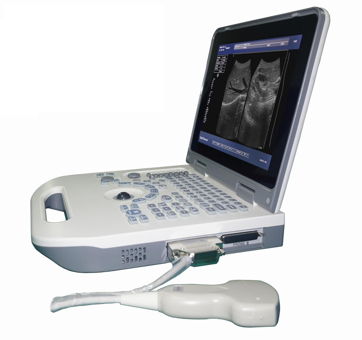
Health care workers who perform ultrasound examinations always apply a layer of clear gel before touching the probe with the examiner. You might think it’s a lubricant, but that’s just a secondary role. The official name of this gel is Ultrasonic Coupling Agent, and its main purpose, as its name suggests, is as a medium for “coupling”. Ultrasonography scans the human body with ultrasound waves (frequency greater than 20 kHz) that are inaudible to the human ear, and converts the echo signals into image information inside the body. B-ultrasound presents black and white images with different brightness (Brightness), while color Doppler ultrasound presents the blood flow of tissues in color on this basis. However, as a mechanical wave, the propagation of sound waves is affected by changes in the medium. Although the probe of the ultrasound equipment is next to the skin, the dry contact is inevitably separated by air. Due to the large difference in acoustic impedance between the air and the human body, two completely different media, sound waves will be reflected a lot at the interface between the two, and a lot of sound energy has been lost before entering the human body. In order for sound...
-
What Is Doppler Ultrasound?
Doppler ultrasound is a non-invasive test that uses high-frequency sound waves (ultrasound) reflected from circulating red blood cells to assess blood flow in blood vessels. Conventional ultrasound uses sound waves to create images but cannot show blood flow. Doppler ultrasound can help diagnose many diseases, including: Thrombus Poorly functioning valves in the veins of the legs, which may cause blood or other fluids to build up in the legs (venous insufficiency) Heart valve defects and congenital heart disease Arterial blockage (arterial occlusion) Reduced blood circulation in the legs (peripheral arterial disease) Arterial bulge (aneurysm) Narrowing of arteries, such as in the neck (carotid stenosis) Doppler ultrasound can assess the speed of blood flow by measuring the rate of change in pitch (frequency). During a Doppler ultrasound examination, a technician (sonographer) trained in ultrasound imaging presses a small, soap-sized hand-held device (transducer) against the skin of the area being examined and removes it from an area as needed Move to another area. This test is an alternative to more invasive tests such as angiography, which involves injecting a contrast agent into the blood vessels so that they can be seen clearly on X-ray images. Doppler ultrasonography can also help doctors check...
-
feilvbin
-
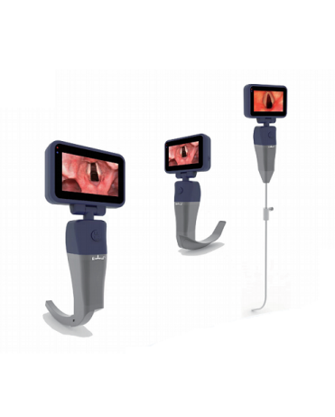
Advantages Of Video Laryngoscope
1. Advantages of video laryngoscope There is no doubt that videolaryngoscopy has been one of the major advances in anesthesia practice in recent years. The main challenge is to determine to what extent videolaryngoscopy should be used in routine clinical practice and which videolaryngoscope will perform best. Going from a standard Macintosh laryngoscope to a video laryngoscope is like going from a standard mobile phone to a smartphone. There have been calls for videolaryngoscopy as a first-line tool for airway management. Most importantly, in the 2015 guidelines of the British Difficult Airway Association, the role of videolaryngoscopy in the management of difficult tracheal intubation has been recognized. The guideline recommends that all anesthesiologists should be trained in the use of videolaryngoscopes and have immediate access to videolaryngoscopes at any time. Furthermore, videolaryngoscopy has been recommended for endotracheal intubation in obese patients, who are known to be at higher risk of airway management-related complications. Beyond the field of anesthesia, it has been predicted that videolaryngoscopy will dominate the field of emergency airway management in the future. Price appears to be the main factor holding back the tide for now. The main reasons that make people enthusiastic about video laryngoscopy are as...
-
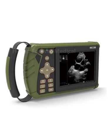
The veterinary B-ultrasound system optimizes and controls some modules
The video signal, video system, and image size of the veterinary B-ultrasound system are optional. Users who use the veterinary B-ultrasound system can freely choose the video signal and video system image size according to different needs to ensure that the program and more capture cards compatibility. The veterinary B-ultrasound system is free to set the basic information. Users can freely fill in or change the name of the animal hospital, clinical diagnosis template and other information. The application for animal doctors can be automatically associated, and it supports manual changes. The veterinary B-ultrasound diagnosis corresponds to the template, that is, after selecting the corresponding diagnosis, the program will automatically call the corresponding template for the user to use. The veterinary B-ultrasound system can be set freely and flexibly with multi-video source interface control. The veterinary B-ultrasound system should require that the workstation can access multiple video signals, and can switch video sources in real time during operation, realizing “one machine with multiple functions”. Veterinary B-ultrasound system prints personalized settings, users can freely set the content of the header and footer of the printed report, and add other information, such as the contact number of the department, which...
-
Effect of B-Ultrasound on Cattle in Preventing Termination of Pregnancy in Dairy Cows
Reproductive obstacles caused by extensive management and overuse of dairy cows in production: due to extensive management, no lactation period, dry milk period, even regardless of size, mixed group feeding under the same ration formula, resulting in poor body condition of lactating cows, cattle The backfat thickness of lactating cows is poorly checked with B-ultrasound. Breeding cows and dry dairy cows are too obese when examined by B-ultrasound, resulting in low reproductive function of the whole herd of cattle. Excessive utilization and blind pursuit of high milk yield lead to reproductive obstacles in dairy cows. There are cases in which milk production is increased by increasing the supply of high-protein concentrate feed. The result of this blind pursuit of milk yield can only cause reproductive disorders and digestive problems. Systemic diseases occur frequently. Reproductive obstacles caused by improper breeding techniques of dairy cows: estrus observation time is wrong, insemination cannot be timely, and methods such as monitoring follicles with B-ultrasound are not used; artificial insemination personnel are not skilled in artificial insemination, artificially causing cervical Injury; lax aseptic operation, retained placenta failed to deal with in time, resulting in endometritis; Vaginal prolapse, etc. cannot be diagnosed and treated in time,...
If you have any question, please contact us