-

Diagnosis of several gastrointestinal diseases in sheep with B-ultrasound
Belly distension of sheep: The left skin of sick sheep is rapidly swollen due to excessive gas in the rumen, with drum sounds on percussion, strong elasticity on palpation, dull spirit, loss of appetite, restlessness in rising and lying, arching the back and moaning, looking back at the abdomen. In severe cases, the breathing speeds up and death from exhaustion occurs. When diagnosing this disease, it is also possible to use B-ultrasound to check the rumen status of sheep for auxiliary diagnosis. Prevention: Feed less fermentable succulent feed to prevent sheep from eating spoiled or poisonous forage and poisonous insects, and often use B-ultrasound to check the rumen status of sheep. Treatment: rumen massage, gavage with anti-fermentation agent, rumen puncture to exhaust gas, and rumen peristalsis drugs attached. B-ultrasound examination for sheep can be used after treatment. Abdominal relaxation in sheep: caused by decreased peristaltic function of the anterior stomach, clinically manifested as indigestion. The cause of the disease is long-term feeding of poor quality and indigestible forage, or too much concentrate and too little grass, which makes the gastrointestinal burden too heavy to adapt to, and even insufficient drinking water, improper feeding methods, and mutations in forage can also...
-
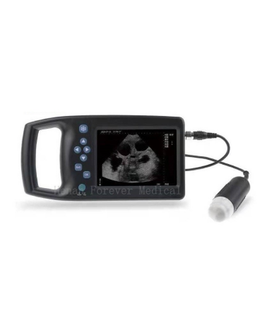
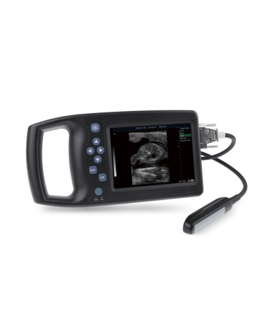
Knowledge of veterinary B-ultrasound probe
A veterinary ultrasound probe is a device that converts one form of energy into another. In ultrasound, sound waves are generated by electrical charges, causing a piezoelectric disk inside the probe to vibrate. Piezo is derived from the Greek word meaning “to press”. Piezoelectric materials are piezoelectric materials, such as quartz, that change shape and size when exposed to an electric field. Piezoelectric crystals are usually cylindrical, 1 to 2 cm wide and 1 mm thick. The current causes the crystal to expand and contract depending on the polarity of the current, and this vibration produces sound waves. The crystals also act as receivers, converting reflected sound waves into electricity, which in turn is converted into modified radio waves that produce images on the screen. Other piezoelectric substances include barium titanate, lithium sulfate, and lead zirconate. Barium titanate is most commonly used. Sensors of various frequencies produce different penetrations. The frequency range is 1-25 MHz; frequencies of 3.5, 5.0, and 7.5 MHz are most common. High frequency probes produce short high frequency wavelengths (i.e. 7.5 MHz) and scan shorter distances with sharper resolution, low frequency probes produce longer wavelengths with greater beam penetration. Although these probes can scan at greater...
-
What you need to know about ultrasonography?
Abdominal ultrasonography: Abdominal ultrasound is the most common, usually what we call liver, gallbladder, pancreas, and spleen examinations. Applicable to: Abdominal screening for abdominal pain, bloating, abdominal discomfort, etc., and inspections for certain organ diseases. Requirements: Fasting for more than 8 hours. Fasting means not eating or drinking anything. I often encounter patients who say that they have not eaten, and if they ask again, they will tell you that they drank some milk or a bowl of soy milk, which is not acceptable. (Of course, if you need to take medicine, you can drink a little water). Urinary tract ultrasonography: Ultrasound of the urinary system includes examination of the kidneys, ureters, bladder, and prostate in males. It is mainly suitable for the screening of tumors, stones, abnormal development of the urinary system, and abnormal urine routine findings. Requirements: Simple kidney examination does not need to be prepared. The bladder, ureter, and prostate need to be filled. You can drink 1000 to 1500 ml of water (or various beverages) 1 to 2 hours before the examination, and hold back the urine after drinking the water. It is difficult to quantify the degree of holding back the urine. It feels the...
-
Do you know these knowledge about ultrasonography?
1. Is ultrasound examination harmful to the body? The medium used in ultrasonic examination is ultrasonic waves, which have no ionizing radiation. The conduction in the human body utilizes the characteristics of sound waves, just like our speaking voice. Ultrasound examination is a very safe examination, so it is commonly used in obstetrics for fetal examination. 2. Is color ultrasound more advanced than B ultrasound? The full name of color ultrasound is color Doppler ultrasound. B ultrasound is B-type two-dimensional ultrasound. Color ultrasound is based on B-type two-dimensional ultrasound with the addition of color Doppler inspection function. It can check the blood supply of organs and blood flow spectrum. Whether the shape and blood flow velocity are normal, so color Doppler ultrasound is more widely used in clinical practice. 3. What is the substance applied during the ultrasound examination? During an ultrasound examination, the doctor will apply a layer of jelly-like substance on the examination site. It is called a coupling agent, which is a water-soluble liquid that isolates the air and allows the ultrasound probe to be in better contact with the skin. The conduction of sound waves ensures the inspection effect, without any poisonous effect, just wipe it...
-
How much do you know about ultrasonography?
1. Which parts of the ultrasound examination need fasting? Ultrasonic examination of the liver, gallbladder, pancreas, adrenal gland, renal artery, left renal vein (nutcracker), etc. requires fasting for 8 to 12 hours. 2. Why fasting? This is determined by the inherent acoustic characteristics of ultrasound, which is afraid of interference from intestinal gas. Because after eating, the gas in the gastrointestinal tract will increase, which will affect the imaging effect, and the doctor will not be able to see clearly. In addition, after eating, the gallbladder shrinks, and the lesions in the gallbladder cannot be detected in time. 3. Which parts of the ultrasound examination need to hold back urine? Examination of uterine appendages, urinary system, prostate, seminal vesicles, etc. requires moderate holding back of urine. 4. Why do you need to hold back your urine? After holding back the urine moderately, the bladder can serve as a good sound-transmitting window, and can push the intestines away, so that the image will be clearer. However, excessive holding back of urine may cause difficulties in diagnosis. Excessive holding back of urine will push the uterus backward, which is not conducive to observation. Note: After 3 months of pregnancy, you can check...
-
Ultrasonic detection of dogs by veterinary B-ultrasound
c Veterinary ultrasound equipment directs a narrow beam of high-frequency sound waves to an area of interest. Sound waves can be transmitted, reflected or absorbed through the tissue they encounter. Are there any downsides to this technology? Ultrasound does not pass through air. Veterinary B-mode ultrasonography is of little value in examining organs that contain air. Ultrasound does not pass through the air, so it cannot be used to examine normal lungs. Bones also block ultrasound, so the brain and spinal cord can’t be seen with ultrasound, and obviously bones can’t be examined. Are there different forms of ultrasound? Ultrasound can take various forms depending on the images produced. Among B-mode (brightness-mode) ultrasound in veterinary work, commonly referred to as two-dimensional ultrasound, is the most common form. This gives a two-dimensional image of the scanned organ. This is a type of ultrasound used to examine abdominal structures, make pregnancy diagnoses, assess heart function, and check the eyes for certain eye conditions. Abdominal structures are examined, a pregnancy diagnosis is made, heart function is assessed, and eyes are examined. M-mode (motion mode) is a B-mode in which the motion trajectory of the structure being scanned is displayed. A combination of M-mode...
If you have any question, please contact us