-
Introduction to Imaging—Basic Knowledge of Ultrasound
(1) Basic principles of ultrasound imaging The vibration frequency of the sound source commonly used in ultrasonic diagnosis is 2.5~5.0MHz. When the ultrasonic wave propagates in the human body and passes through different organs, tissues, and lesions, the acoustic impedance of the medium on both sides of each interface is different, and different degrees of reflection, scattering, and ultrasonic attenuation occur. The information is obtained by the receiver, and after information processing, it is displayed as a waveform or image on the screen. According to the type and display mode of ultrasound imaging, it is divided into A-type ultrasound, B-type ultrasound (two-dimensional ultrasound), M-type ultrasound, and D-type ultrasound. (2) Main features of 2D sonogram ①The imaging record is a two-dimensional cross-sectional view of the inspection site, and the inspection site and scanning direction are often marked. ②The image is composed of black, white, and gray dots, which represent the strength of the tissue structure echo, and the whiter the color, the stronger the echo (distinguishing between high-density and low-density X-ray and CT). ③The image display range is limited, and larger organs and lesions cannot be displayed as a whole. ④Contrast-enhanced acoustic examination changes the echo of the tissue structure on...
-

The Safety of B/W ultrasound Scanner
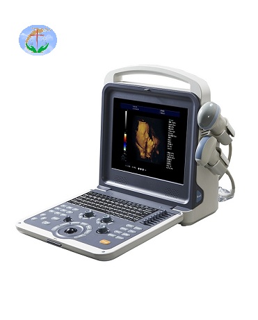
B/W ultrasound Scanner to exceed what damage to the fetus whether, in the medical field, it has not yet authoritative, is the debate, most scholars think B ultrasonic examination to the fetus has no sure of damage. From B ultrasonic to exceed the principle analysis, B ultrasonic ultrasonic transmission is, there is no ionizing radiation and electromagnetic radiation, is a sound wave transmission, the sound waves to the human body organization is not anything to hurt. But if the sound waves in a fixed place dense, and gathered together a long time, it will be a heating, the heating effect to a certain extent, to human tissue may have a bad effect, the influence of the cell substances, including chromosomes. Theory is of high strength of ultrasonic can through its high temperature and the role of the organizations cavity, damage to the organization. But in fact, the medical use of B ultrasonic super is low intensity, less than 94 mw/cubic centimeter, it is not a risk to the fetus, is still not B ultrasonic ultrasonic examination cause fetal malformation reports. At present, all the hospitals in a maternity used in the field of fetal B ultrasonic examination is safe. But...
-
Popular science knowledge of ultrasonography
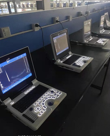
Ultrasound is to use the ultrasonic probe to repeatedly scan and receive the ultrasonic sound beam, and display the received different echo intensities on the corresponding position of the screen to outline the gray-scale section image. The received ultrasound is a high-frequency sound wave, which is safe and harmless to the human body. It is also very safe to be widely used in the examination of various organs of the body and the fetus. Among them, interventional ultrasound is also a minimally invasive treatment. Under the real-time dynamic monitoring and guidance of ultrasound, various operations such as puncture, biopsy, suction, intubation, drug therapy, microwave, and radiofrequency ablation are completed. The operation time is short, recovery is fast, It has the advantages of fewer complications and has been widely used in clinical practice. Do Ultrasounds Have Radiation? The “ultra” in ultrasonography refers to ultrasound, which is essentially the same as the sound you hear, but ultrasound is a special high-frequency sound wave that cannot be heard by the human ear, so there is no radioactivity, so you can check it with confidence. The advantages of ultrasonography, such as economy, convenience, and repeatability, have now become one of the first-choice inspection methods...
If you have any question, please contact us